Magnetic resonance imaging
What is MRI?
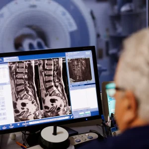
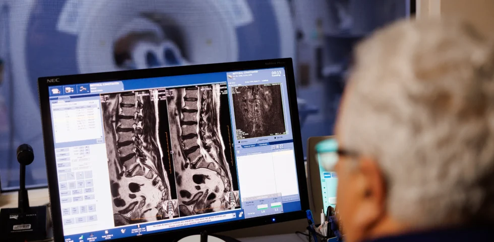
Magnetic Resonance Imaging (MRI) is one of the most advanced diagnostic imaging methods currently available. It allows detailed visualization of the organs and tissues of the body, helping to establish an accurate diagnosis and choose the appropriate treatment. Unlike computed tomography (CT) and radiography, MRI does not use ionizing radiation, but a strong magnetic field and radio waves to generate high-resolution images.
What are the advantages of the investigation?
MRI offers numerous benefits:
- Detailed images of organs and soft tissues;
- Visualization of anatomical structures in multiple planes, without repositioning the patient;
- Accurate assessment of the brain, spine, joints, abdominal and pelvic organs;
- Examination of blood vessels, sometimes without the administration of contrast material;
- The possibility of using advanced techniques that provide information about both the structure and function of tissues;
- Lack of exposure to ionizing radiation.
How is the investigation conducted?
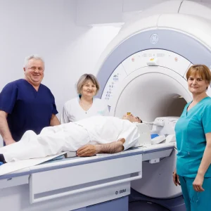
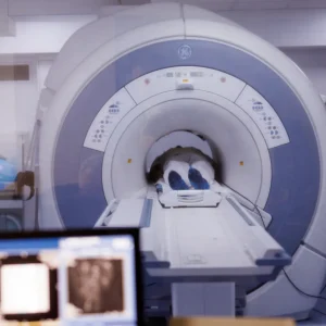
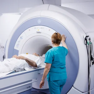
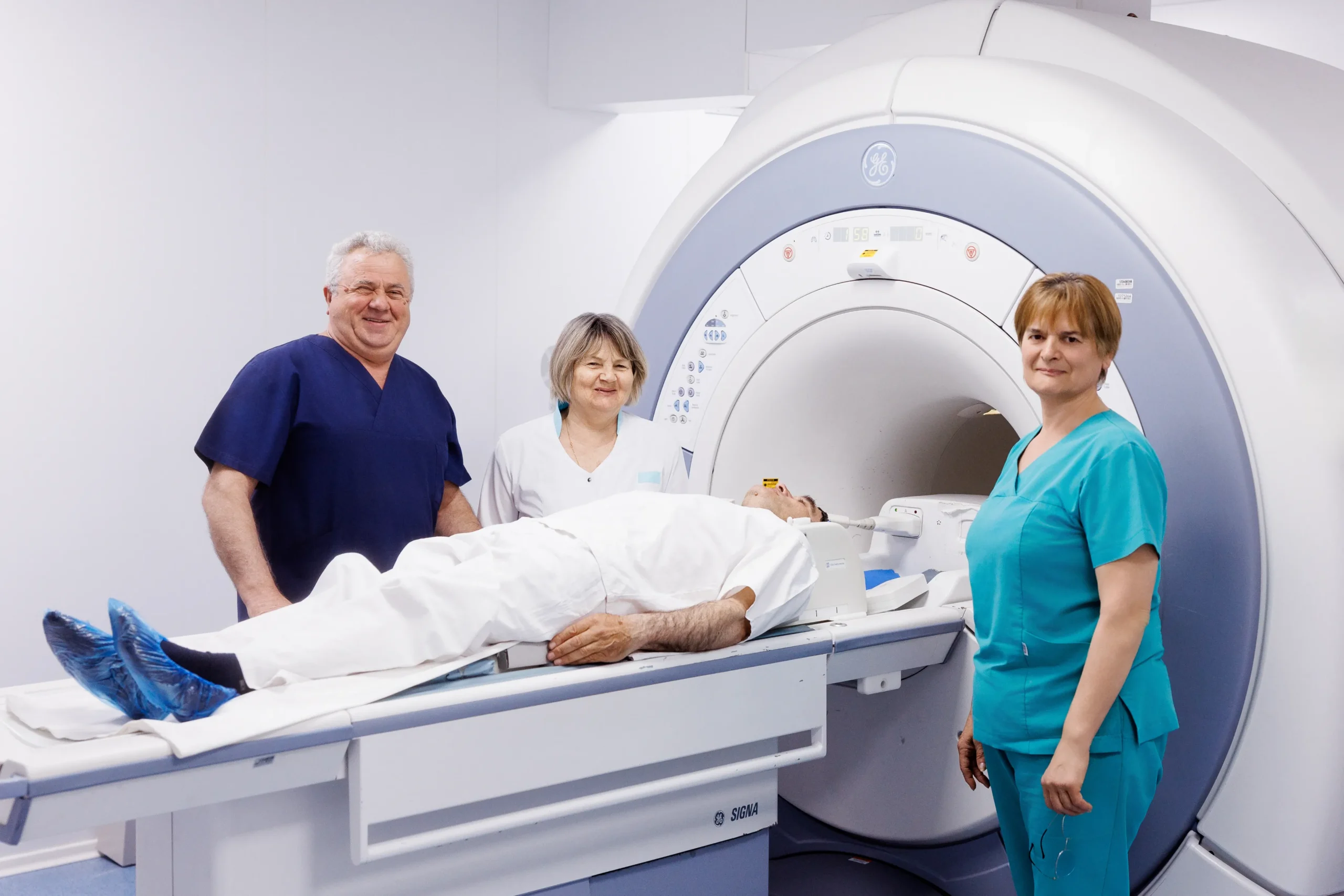
The patient is placed on a movable table that is inserted into the MRI machine, a cylindrical tunnel containing a powerful magnet. The duration of the examination is generally between 30 and 60 minutes, depending on the area examined and the protocol used. During the scan, the machine produces loud noises, which is why the patient is given headphones or earplugs. To obtain quality images, it is important that the patient remains still throughout the examination.
What conditions and areas of the body can be investigated?
MRI is used to diagnose and monitor a wide range of conditions, including:
- Pathologies of the brain, spinal cord and nerves;
- Spine and joint diseases;
- Tumors and other structural changes in internal organs;
- Diseases of the liver, biliary tract and abdominal organs;
- Gynecological conditions, including endometriosis and uterine fibroids;
- Pathologies of the prostate and other pelvic organs;
- Evaluation and monitoring of certain forms of cancer.
Is it necessary to administer contrast material?
In certain situations, to increase the accuracy of the diagnosis, a gadolinium-based contrast agent may be administered. The need for its use is determined by the attending physician or radiologist, depending on the medical indication and the images obtained.
Is this a safe investigation?
MRI is a painless, minimally invasive procedure and considered very safe. Discomfort can only be caused by the noise of the machine, the confined space or the administration of contrast material, when necessary. Since it does not use ionizing radiation, the investigation can be repeated whenever necessary, including to monitor the evolution of a condition.
Is special training required?
In most cases, MRI examination does not require any special preparation. However, before the examination, the patient must remove all metal objects, including jewelry, watches, and clothing accessories that contain metal.
It is important that the medical staff is informed about the existence of implants, prostheses or other medical devices. Depending on the area examined, the doctor may recommend certain instructions regarding diet, fluid consumption or administration of certain medications before the investigation.
MRI and pregnancy
If you are pregnant or suspect you are pregnant, inform your doctor before the examination. MRI examinations are generally avoided in the first trimester, except when absolutely necessary, and the administration of contrast material is not recommended during pregnancy.
MRI is one of the most accurate and safe modern imaging methods, providing detailed information about the internal structures of the body without exposure to radiation. Due to its high accuracy, this investigation plays an essential role in the diagnosis and monitoring of numerous conditions.