Radiology and computed tomography
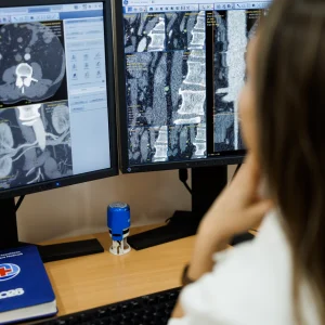
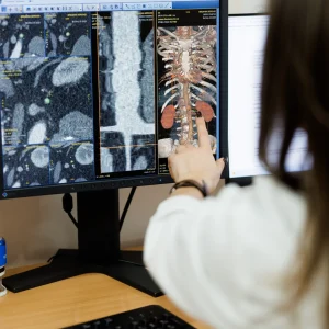
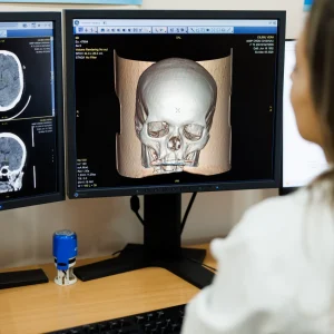
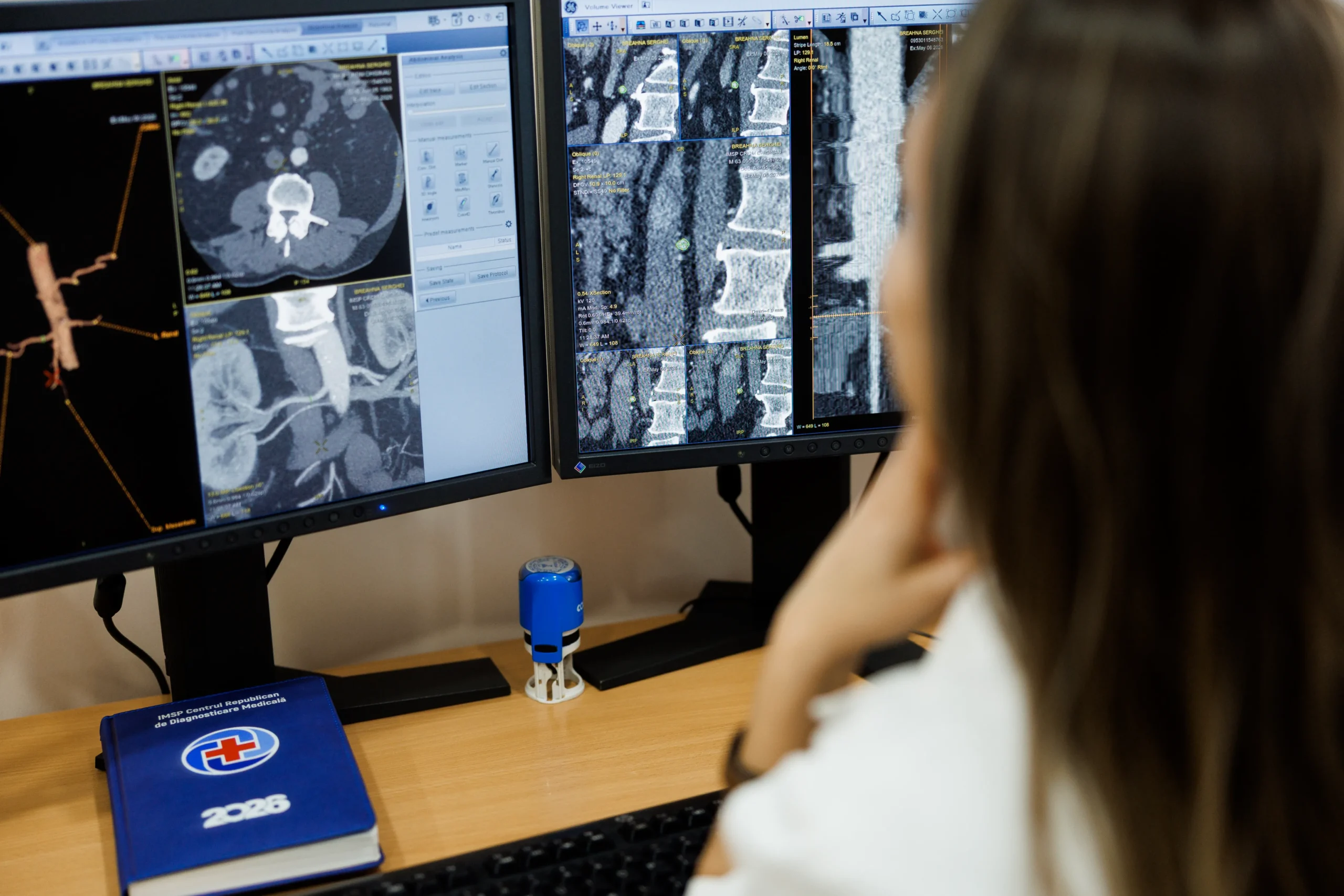
Computed tomography (CT) is a modern medical imaging method that uses X-rays and computer technology to obtain detailed images of internal organs, bones, and blood vessels. The investigation allows visualization of body structures in thin sections ("slices"), providing the doctor with precise information for diagnosis and treatment monitoring. In certain situations, the examination is performed with the administration of iodine-based contrast material, to better highlight organs and vessels. CT is a quick, painless procedure and very useful in emergencies or in the detailed evaluation of various conditions.
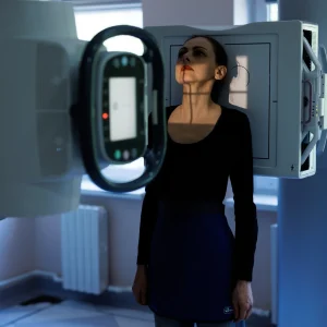
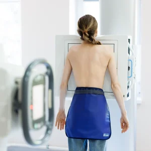
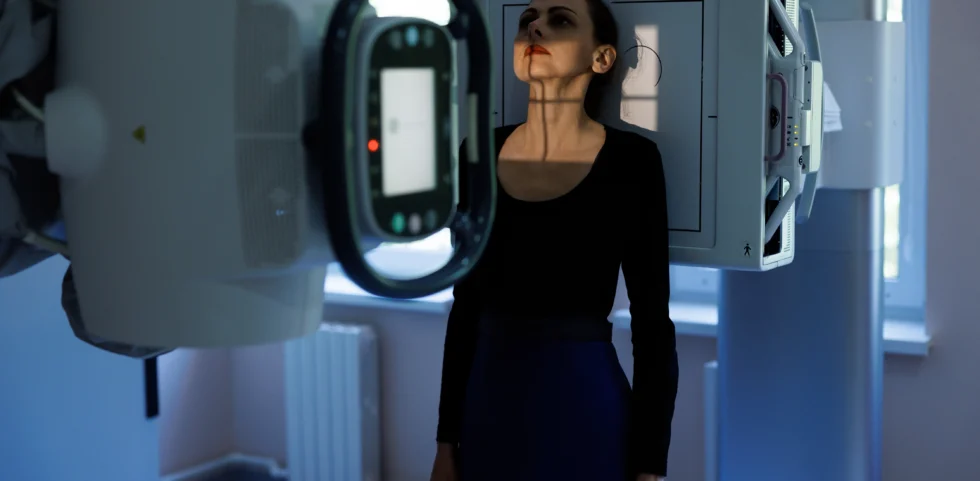
Radiography (X-ray) is one of the most common and accessible imaging tests, which uses X-rays to visualize the internal structures of the body, especially the bones and organs of the chest. It is used to diagnose fractures, lung diseases (such as pneumonia), and to evaluate changes in the joints or spine. X-rays are a quick, painless procedure and involve a low dose of radiation.
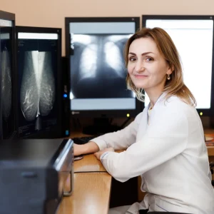
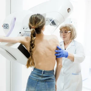
Mammography is a specialized imaging test that uses low doses of radiation to examine the breasts. It is the most effective method for early detection of breast cancer, being able to identify very small changes, even before they are palpable.
Mammography is recommended periodically, especially for women over 40 or as directed by a doctor. The procedure takes a few minutes and may cause slight temporary discomfort due to the compression of the breast, which is necessary to obtain quality images.
The following radiological examinations are performed in the department:
- Mammography of the mammary glands;
- Chest X-ray;
- Abdominal cavity radiography;
- Urography;
- Skull radiography;
- X-ray of the orbits;
- X-ray of the clavicle;
- X-ray of the paranasal sinuses;
- Bone X-ray;
- C1 radiograph through open mouth;
- X-ray of the sacral bone;
- Hip joint radiography;
- X-ray of the pelvic bones;
- Hand radiography;
- X-ray of the knee joint;
- Computed tomography of the brain without contrast;
- Computed tomography of the brain with contrast;
- Computed tomography of the abdomen;
- Computed tomography of the pelvis;
- Computed tomography angiography of the arteries;
Toată lista de investigații:
Mammography of the mammary glands, Chest radiography, Abdominal cavity radiography, Urography, Skull radiography, Orbital radiography, Clavicle radiography, Paranasal sinus radiography, Turkish saddle radiography, Bone radiography, C1 open-mouth radiography, Cervical/thoracic/lumbar spine radiography, Sacral bone radiography, Hip joint radiography, Pelvic bone radiography, Humeral joint radiography, Arm bone radiography, Forearm bones radiography, Elbow joint radiography, Radiocarpal joint radiography, Hand radiography, Femur radiography, Knee joint radiography, Calf radiography, Talocrural joint radiography, Plantar radiography, Sacroiliac joint radiography, Calcaneal radiography, Flatfoot radiography (bilateral), Non-contrast brain computed tomography, Contrast-enhanced brain computed tomography, Chest and mediastinal computed tomography without contrast, CT scan of the chest and mediastinum with contrast, CT scan of the abdomen without contrast, CT scan of the abdomen with contrast, CT scan of the pelvis without contrast, CT scan of the pelvis with contrast, CT scan of the paranasal sinuses, CT scan of the paranasal sinuses and brain, CT scan of the nasopharynx, CT scan of the nasopharynx with contrast, CT scan of the thyroid gland, CT scan of the thyroid gland with contrast, CT scan of the vertebrae, CT scan of the joints or knee, CT scan of the femur (calf), CT scan of the talocrural joints, CT scan of the temporal bones, CT scan of the orbits, CT scan of the abdomen and pelvis in the urographic phase, Duplication of the CT scan result of an area/organ/compartment, Spiral CT scan with three-dimensional image - virtual bronchography, CT scan biphasic CT of the liver, pancreas with contrast, Triphasic CT of the liver, pancreas with contrast, Oncological assessment CT (thorax, abdomen + pelvis) without contrast, Oncological assessment CT (thorax without contrast, abdomen + pelvis with contrast), Aortography, Computed tomography angiography of the arteries of the pelvis, Computed tomography angiography of the coronary arteries and ventricles, Computed tomography angiography of the carotid arteries, Computed tomography angiography of the cerebral arteries, Computed tomography angiography of the hepatic and pancreatic arteries, Computed tomography angiography of the pulmonary arteries, Computed tomography angiography of the limb arteries, Computed tomography angiography of the renal arteries.
Recommendations for patient preparation:
General recommendations:
Inform your doctor if you are pregnant or suspect pregnancy
Presentation with medical referral and relevant previous documentation
Informing the patient about the procedure
Removal of metal objects from the examined area
Evaluation of allergies, especially to iodinated contrast agents
For CT examinations without contrast:
Usually does not require special preparation
Avoiding heavy eating before the examination (optional)
For X-ray investigations:
Usually no special preparation is required
Follows staff directions (e.g., holding a position or holding your breath for a few seconds)
For CT or X-ray examinations with contrast:
Fasting 4–6 hours before the examination
Adequate hydration before and after the procedure
Renal function assessment (UREA AND SERUM CREATINE), with results no older than 2 weeks
Information regarding any previous allergic reactions
Avoiding taking Metformin as recommended by your doctor (if applicable)
For abdominal examinations:
Light diet 24 hours before
Avoiding foods that produce gas
Possibly administration of oral contrast material (as directed by the doctor)
For pelvic examinations:
Moderately full bladder (in some cases)
For mammography:
Schedule the investigation, preferably, in the first part of the menstrual cycle (for increased comfort)
Do not use deodorant, lotions or powder in the armpit and breast area on the day of the examination
Wear comfortable, two-piece clothing (to facilitate the examination)
Present previous mammograms or ultrasounds (if any)
Inform your doctor about any symptoms or changes you notice in your breasts