
Imaging investigations: CT, MRI and ultrasound
When pain, persistent symptoms, or changes occur that require evaluation, your doctor may recommend imaging tests to examine the internal structures of the body. These tests allow visualization of organs, tissues, bones, and blood vessels, providing important information for establishing a diagnosis. At the Republican Center for Medical Diagnostics, patients can benefit from computed tomography, magnetic resonance imaging, and general ultrasonography, depending on the doctor's recommendation and the particularities of each case.
What role do imaging investigations play?
Sometimes, symptoms are not enough to determine the exact cause of a condition. Abdominal pain, dizziness, trauma, inflammation, or certain changes observed on other investigations may require additional imaging examinations. These investigations can help identify inflammation, injury, or other internal changes and allow evaluation of abdominal, thoracic, and pelvic organs, as well as the brain, spine, joints, and soft tissues. They are also useful for monitoring the progression of conditions and evaluating the results of treatment.
Computed tomography (CT)
Computed tomography (CT) is an imaging test that uses X-rays to produce detailed images of different areas of the body. It may be recommended in the evaluation of trauma, persistent pain, pulmonary, abdominal, or neurological changes. CT is considered a quick, painless and non-invasive investigation, and in certain situations it can be performed with contrast material for more detailed visualization.
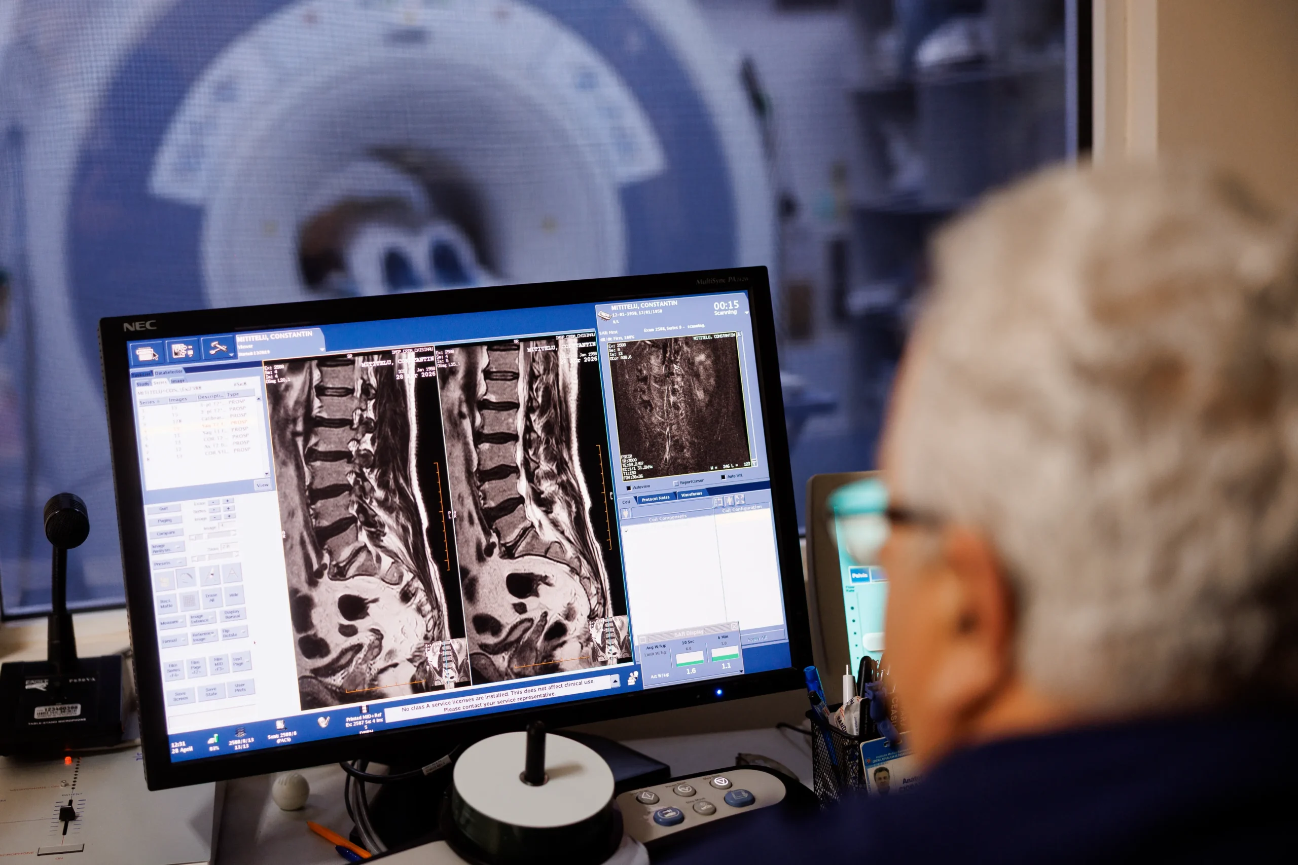
Magnetic resonance imaging (MRI)
MRI uses magnetic fields and radio waves to produce detailed images of internal structures without the use of X-rays. This examination may be recommended for evaluating the brain, spine, joints, soft tissues, and internal organs. MRI is useful when a detailed analysis of certain structures or monitoring of the evolution of certain conditions is necessary. Depending on the medical indication, the investigation can also be performed with contrast material.
Ultrasound
Ultrasound, also called ultrasonography, is an imaging investigation performed with ultrasound and does not use X-rays. It allows the examination of various internal organs and tissues and is frequently used in diagnosis and monitoring. Ultrasound may be recommended to evaluate the abdominal organs, urinary system, thyroid gland, mammary glands, reproductive system, or blood vessels. It is also commonly used to investigate pain, inflammation, and changes in blood circulation.
How is the appropriate investigation determined?
CT, MRI, and ultrasound have different roles and are recommended depending on the symptoms, the area examined, and the information needed for diagnosis. CT may be indicated when rapid and detailed images are needed, MRI is frequently used to evaluate soft tissues and the nervous system, and ultrasound is particularly recommended for abdominal, gynecological, breast, or vascular examinations.
The doctor is the one who determines the appropriate investigation for each patient.
Preparing for investigations
Preparation varies depending on the type of investigation and the area being examined. In some cases, the patient may receive recommendations regarding diet, fluid intake, medication administration, or additional tests. It is important that the doctor is informed about allergies, known conditions, treatments administered, pregnancy or implanted devices, especially before contrast material examinations or before MRI.
Imaging investigations at CRDM
Imaging investigations contribute to the identification of changes that cannot be observed only through consultation or laboratory analysis and play an important role in establishing the diagnosis. At the Republican Center for Medical Diagnostics, patients can benefit from CT, MRI, and ultrasound, depending on medical recommendation and the needs of each case.